

User Manual
Features
-
- Assisted use with Dr. Ann Fu or unassisted use after training.
- Applications include single region or cell isolations for extraction of DNA, RNA, protein or metabolites from nearly any biological system, allowing downstream (cell-specific) analyses such as PCR, RNA-sequencing, qRT-PCR, microarray, genetic fingerprinting, 2-D protein gels, and mass-spec analysis (LC-MS/MS/MALDI). See Leica science lab for more information.
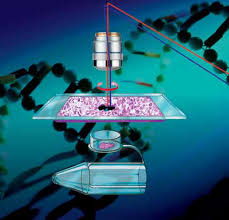
- Fully automated high-end upright microscope.
- Dedicated LMD objectives available: 5x, 10x, 20x, and 40x.
- Special membrane-coated (PEN) glass or frame slides are recommended for mounting tissue sections.
- Three-slide holder with rapid 2.5x slide scans for over-view images.
- Fully integrated fluorescence allows live cutting within bright-field and fluorescence (BGR filters for DAPI, FITC/GFP, TRITC/PI) mode.
- UV laser 349 nm solid state diode (120 µJ, pulse frequency 10 – 5000 Hz, pulse length < 4 ns), with continuously adjustable laser aperture, speed, and focus.
- Gravity collection into standard PCR microfuge caps (0.2, 0.5ml), 8-strip wells, or cut live cells in Petri dishes with or without special membrane (Leica or Ibidi slides).
- Annotate then batch cut or cut in real-time using the Pen-screen or keyboard mouse.
- Export each object area for tabulation.
- Image data management:
- Please always transfer your data after you finish imaging. Any images left on the computer hard-drive may be deleted to restore hard drive space.
NIH Acknowledgement in Publications
- Please acknowledge funding for this instrument as follows: This work utilized a LEICA 7000 laser microdissection microscope purchased with an NIH shared instrumentation grant 1S10OD016350-01 and operated by the University of Florida Molecular Pathology Core (RRID:SCR_016601).
Publications
- Campbell-Thompson M, Butterworth, E.A., Boatwright, J.L., Nair, M.A., Nasif, L. H., Nasif, K., Revell, A.Y., Riva, A., Mathews, C. E., Gerling, I. C., Schatz, D.A., Atkinson, M.A. Islet sympathetic neuroanatomy in human type 1 diabetes. Scientific Reports, 2021 Mar 22;11(1):6562. PMID: 33753784 PMCID: PMC7985489 DOI: 1038/s41598-021-85659-8
- Dou M, et. al. Nanowell-mediated two-dimensional liquid chromatography enables deep proteome profiling of <1000 mammalian cells. Chemical Science. 2018.
- Zhu Y, et. al. Nanodroplet processing platform for deep and quantitative proteome profiling of 10-100 mammalian cells. Nat Commun. 2018.
- Han S, et al. The pancreatic tumor microenvironment drives changes in miRNA expression that promote cytokine production and inhibit migration by the tumor-associated stroma. Oncotarget, 2016. link
Fees and Invoicing
Please visit the Fee page for information on costs and billing.