

- Histology
- Immunohistochemistry (IHC/IF/ISH)
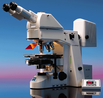
- Brightfield and fluorescence microscopy and whole slide scanning
- Laser microdissection microscopy
- Tissue Microarrays
- Cytospins
- Equipment for rent
- Digital Pathology – Whole Slide Scanning
- Nanostring GeoMX Digital Spatial Profiling
- Nanostring nCounter
First-time submitter?
- Don’t hesitate to get in touch with us to discuss any aspect of your project.
(352) 273-7749
molecular@ufl.edu - Drop off and pick up samples in D11-41 (Dental Tower, 11th floor). Email your submission form.
- You will be notified by email or phone when work is completed and ready for pick up.
- FAQ
Turnaround Time (TAT)
Submissions are handled first come, first served with every attempt to finish work as soon as possible. Do let us know about deadlines so we can try to meet them. Turnaround times depend on the size of your submission (number of blocks or slides), complexity (standard or developmental stains), and current workload.
General TAT:
- Paraffin process and embedding (FFPE): 1-3 days (Samples are processed in batch by species to decrease user costs. All processor reagents are changed based on the number of processing runs, not by the number of blocks/run).
- Sectioning paraffin blocks to unstained sections (FFPE sections): 1-3 days
- H&E stain slide: 0.5 day
- IHC/IF/ISH: Immunolocalization TAT depends on whether the antibody has been validated and used in species and tissue of interest. Contact Ann Fu to discuss your project before harvesting samples or purchasing your antibodies.
Consultations/Grants
Staff members are happy to consult on any aspect of your project as discussions before sample harvesting can lead to better outcomes. We can also provide letters of support and budgetary information for grant applications.
NIH Acknowledgement in Publications
- If your publication included MPC staff assistance or one of our instruments, we would appreciate your acknowledgment with a statement like “(Histology/Microscopy) was performed at the Molecular Pathology Core in the Department of Pathology, Immunology, and Laboratory Medicine at the University of Florida College of Medicine, RRID: SCR_016601.”
- Please acknowledge funding for the Leica laser microdissection microscope as follows: This work utilized a LEICA 7000 laser microdissection microscope purchased with an NIH shared instrumentation grant 1S10OD016350-01 and operated by the University of Florida Molecular Pathology Core (RRID:SCR_016601).